|
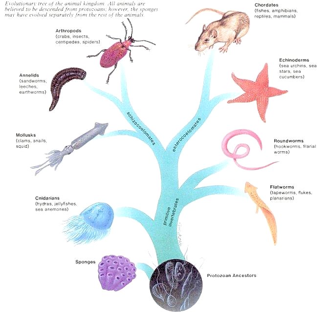
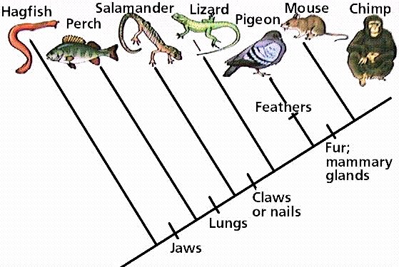
All phyla of animals had evolved by the beginning of the Paleozoic Era some 540 million years ago. The evolutionary tree of animals in Figure 01 indicates that animals are the descendants of protozoans - perhaps in a colonial form whose cells differentiated into various types of cells. The evolutionary tree of animals resembles a tree with 2 main branches. The animal phyla located on the main trunk of the tree are referred to as the primitive invertebrates, and the animals of the main 2 branches include the advanced invertebrates and the vertebrates. Invertebrates lack a dorsal backbone, while vertebrates have a backbone made up of vertebras. A study of the evolution of animals reveals that the most complex animals have the most advanced features as listed in Table 01 below. Classification of animals therefore is based on type of body plan, symmetry, number of |
Figure 01 Evolutionary Tree of Animals [view large image] |
germ layers, level of organization, type of body cavity, and presence or absence of segmentation. |
| Features | Most Primitive | Primitive | Advanced | Most Advanced |
|---|---|---|---|---|
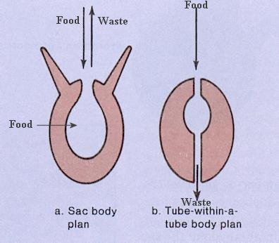
| Body Plan | None | Sac plan | Tube-within-tube plan | + specialization of parts |
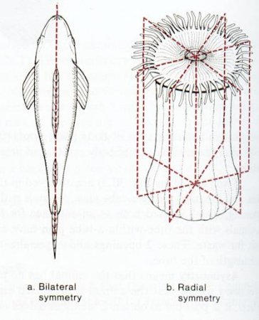
| Symmetry | None | Radial | Bilateral | + cephalization (head) |
| Germ layers | None | 2 | 3 | 3 |
| Level of organization | None | Tissue | Organ | Organ system |
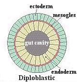
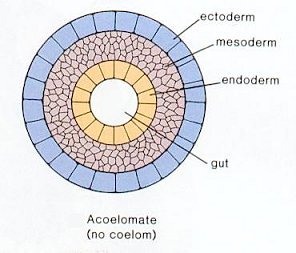
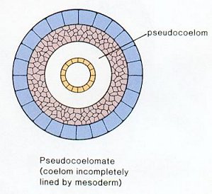
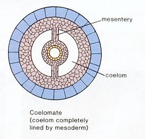
| Body cavity | Diploblastic | Acoelomate | Pseudocoelom | True coelom |
| Segmentation | Nonsegmented | Nonsegmented | Segmented | + specialization of parts |
 |
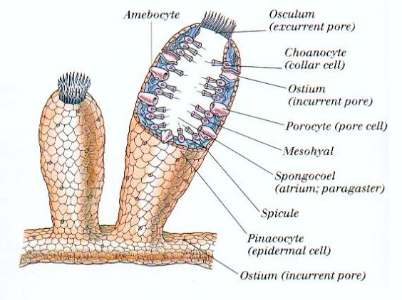 |
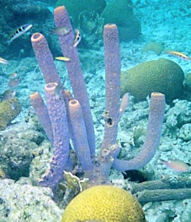
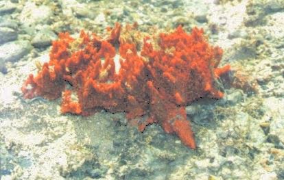
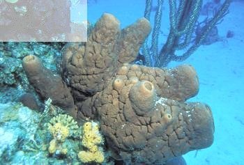
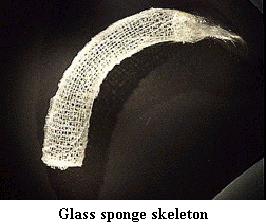
necessary for multicellularity already. Their evolutionary steps are clearly demonstrated from single-celled aquatic protists to colonies and then appear as the collar cells in sponges. A sponge does not have anything in their bodies that can be called tissues or organs. Instead each type of sponge cell is responsible for a different activity to keep the sponge alive. Some sponges grow on rocks and are brightly colored. Sponges often are shaped like vases with a central cavity (Figure 02). Figure 03 shows the anatomy of the sponge. The internal structures are described in the followings. |
Figure 02 Sponge |
Figure 03 Sponge Anatomy [view large image] |
Although the terminologies (and hence the functions) for the various systems are in close parallel to those for the human body, the composition and distribution are vastly different. |
 |
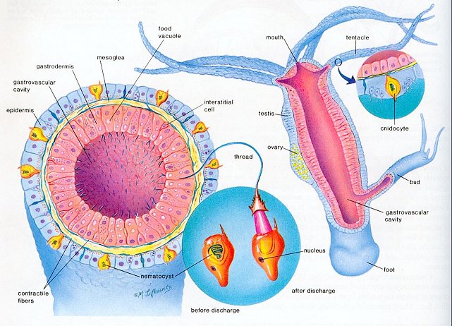 |
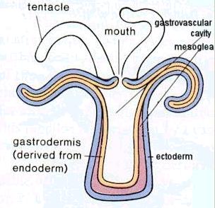
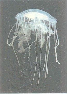
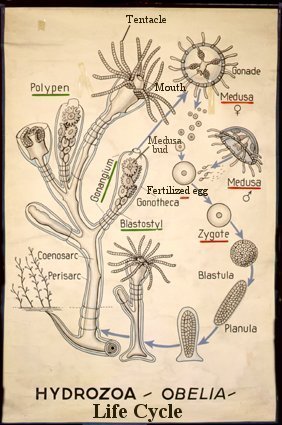
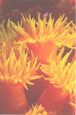
shape with the mouth region directed downward, are called jellyfishes, or medusae. The polyp is adapted to a sessile life, while the medusa is adapted to a floating or free-swimming existence. At one time, both body forms may have been a part of the life cycle of all cnidarians, because today we see an alternation of generations life cycle of these two forms in certain cnidarians, such as members of the genus obelia. In such life cycle, the polyp stage produces medusae, and the medusae, |
Figure 04 Cnidaria [view large image] |
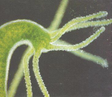
Figure 05 Hydra Anatomy [view large image] |
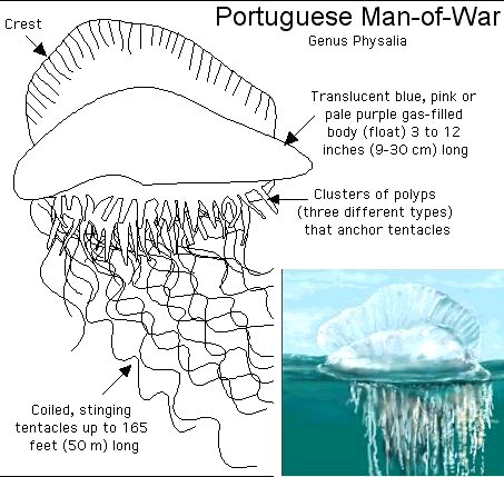
which produce eggs and sperm, disperse the species. Cnidarians are quite diversified including the Portuguese man-of-war, sea anemones, hydra and many other species. |
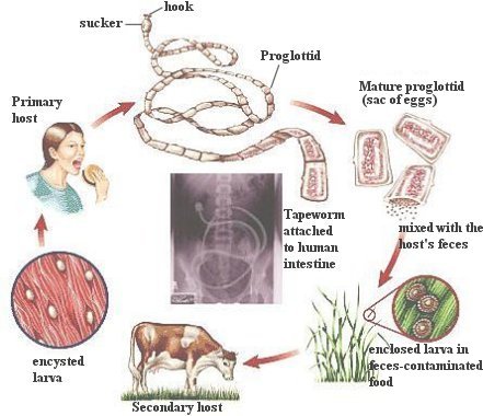 |
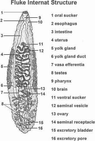
Flatworms are nonsegmented, lack a coelom, and have the sac body plan with only one opening. Therefore, if we analyze them according to Table 01, they have a combination of primitive and advanced features. There are three classes of flatworms: one is free living and two are parasitic. The free-living specimen, the planarian, best exemplifies the characteristics of the phylum. Tapeworms and flukes are parasitic with structure reflecting the modifications that occur in parasitic animals. Concomitant with the loss of predation, there is an absence of cephalization; the anterior end notably carries hooks and/or suckers for attachment to the host. The parasite acquires nutrient molecules from the host, and the digestive system is reduced. It is | Figure 06 Tapeworm Life Cycle [view large image] |
covered by a specialized body wall resistant to host digestive juices. The extensive development of the reproductive system, with the production of millions of eggs, may be associated with difficulties in dispersing the species. Figure 06 shows the life cycle of the tapeworm. |
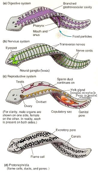 |
| Figure 07 Planarian Anatomy [view large image] |
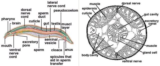 |
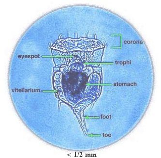
with the rotifers. Roundworms possess two anatomical features not seen in more primitive animals: a tube-within-a-tube body plan and a body cavity. The body cavity is a pseudocoelom, or a cavity incompletely lined with mesoderm. This fluid-filled pseudocoelom provides space for the development of organs, and serves as a type of skeleton. When roundworms are analyzed according to Table 01, they are seen to have features associated with advanced animals except that they are nonsegmented. |
Figure 08 Roundworm Anatomy [view large image] |
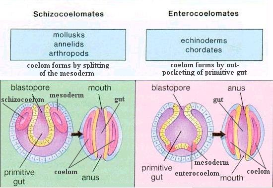 |
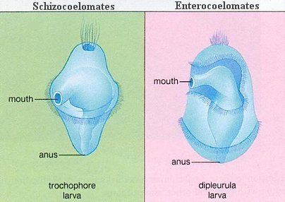
All the advanced invertebrate phyla have a true coelom. Nevertheless, they can be divided into two groups on the basis of embryological evidence (Figure 09). In mollusks, annelids, and arthropods, the coelom forms by splitting of the mesoderm. Therefore, they are called the schizocoelomates. In echinoderms and chordates, the coelom forms by outpocketing of the primitive gut. It is thus called enterocoelomates. Note the interchange of mouth and anus in these two different types of development. |
Figure 09 Coelom Formation |
 |
|
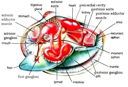
Figure 10 Clam Anatomy |
side of the visceral mass, which lies above the foot. Gills are composed of vascularized, highly convoluted, thin-walled tissue specialized for gas exchange. |
 |
separatly or in groups under stones, logs or fallen leafs. After a few weeks tiny snails are born. They have a transparent shell. Some snailspecies are ovo-viviparious, this means that the eggs hatch inside of the snail. Most species reach maturity in a year, but the larger ones can take two to four years to reach maturity.
|
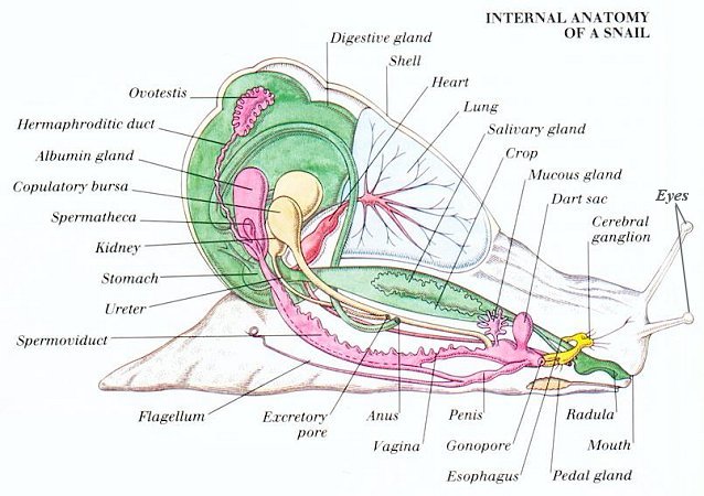
Figure 11 Snail Anatomy |
blood and arranged like the teeth of a comb. |
 |
|
Figure 12 Digestive System |
ink sac to escape danger. They can also change color according to mood and environ- ment, sometimes exhibiting rapid waves of color changes that sweep over the body. |
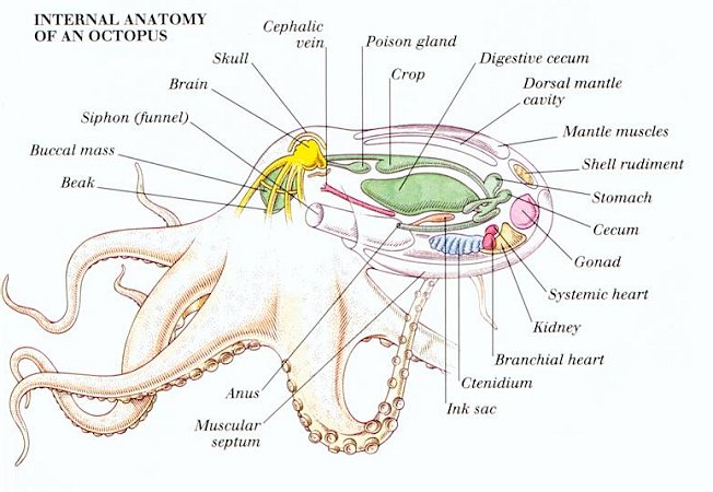 |
same short-term and long-term memories as vertebrate animals.
|
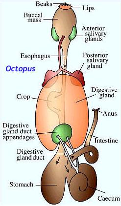
Figure 13 Octopus Anatomy |
Some of the octopus anatomy is shown in Figure 13. |
 |
|
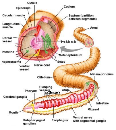
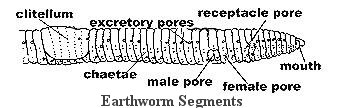
Figure 14 Earthworm Anatomy |
efficient absorption of oxygen dissolved efficient in the water. |
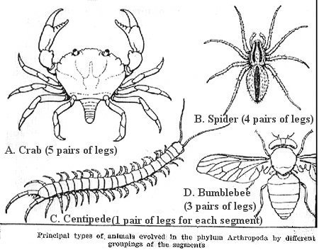 |
Segmented animals have repeating units. This has led to specialization of parts over evolutionary time because the different segments could become specialized for different purposes. The ancestral arthropods probably had many segments with a pair of appendages on each segment. These animals belong to the classes Chilopoda (see Figure 15C) and Diplopoda (not shown, 2 segments fused - 2 pairs of legs for each segment). The segments of arthropods today (classes Arachnida and Insecta, Figure 15B and D) are often fused into regions such as the head, thorax, and abdomen. The head and thorax may be fused to form a cephalothorax (class Crustacea, Figure 15A). Since insects comprise one of the largest animal |
Figure 15 Arthropods |
groups - both in number of species (about 1 million) and in number of individuals, the grasshopper will be used as the specific example. |
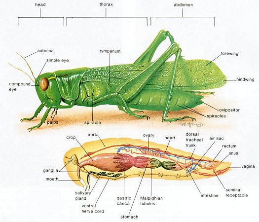 |
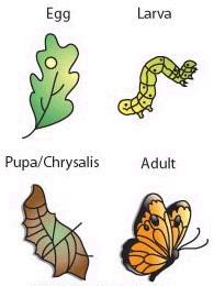
complete metamorphosis. Worm-like larvae form a cocoon called a pupa and then become transformed into the adult form. Flies and butterflies undergo complete metamorphosis. Complete metamorphosis allows the animal to take advantage of two different ecological settings (niches). For example, caterpillars are specialized for feeding on plant leaves. The butterfly of many species is specialized for finding mates and reproduction.
|
Figure 16 Grasshopper Anatomy [view large image] |
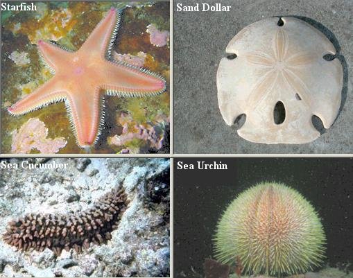 |
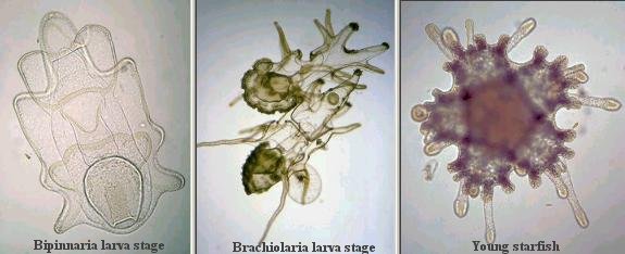
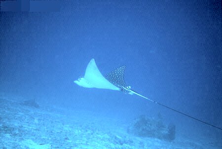
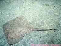
radially symmetrical, in contrast to vertebrates; they have no internal skeleton, no trace of any of the three major chordate characters of notochord, nerve cord, or gill slits, and they have many peculiar and complicated organs of their own. But the embryology sheds an unexpected gleam of light. The early embryo of the echinoderm is a tiny creature, which floats freely in the sea water. Unlike the adult, the larva is bilaterally symmetrical, suggesting that the radial symmetry of the starfish is a secondary affair, assumed when the ancestors of these forms look up a sedentary existence. Then, too, the type of development of certain of the body cavities is identical with that found in the embryos of some primitive vertebrates. It is believed that the bilateral larva developed types which retained the original symmetry, |
Figure 17 Echinoderms |
and gradually evolved into the chordates and, finally, the true vertebrates. |
 |
projecting arms and after some more weeks, a brachiolaria larva is formed. The larvae have their own gut, with inside cilia to inhale and transport food particles. They feed themselves with diatoms and other organisms in the plankton. The stomach is large and round and situated at the backside. After this phase a large part of the larva degenerates and at the rear side a rudimentary formed juvenile starfish develops. The organs of the young starfish are formed anew.
|
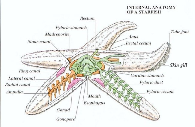
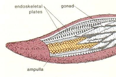
Figure 18 Starfish Anatomy |
function of respiratory exchange. |
 |
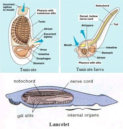 |
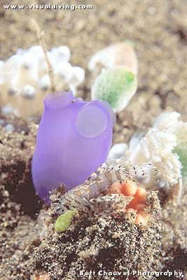
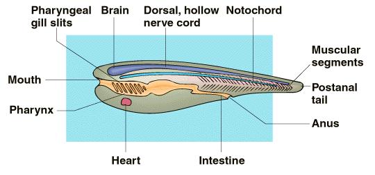
the only chordate feature retained by the adult. The larva of the tunicate, however, has a tadpole shape and possesses the three chordate characteristics. It has been suggested that such a larva may have become sexually mature without developing the other adult tunicate characteristics, and may have evolved into a fishlike vertebrate similar to the lancelet, which is a chordate that shows the three chordate characteristics as an adult. |
Figure 19a Vertebrates |
Figure 19b Protochordates |
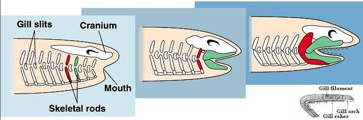 |
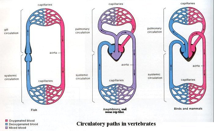
Fishes added a capillary network with gas-permeable walls; this network evolved into gills. It is much reduced in adult land-dwelling forms (although it is extremely important in embryonic development of all vertebrates). Land animals use lungs for gas exchange instead of gills. It is believed that the gill arches have been modified to |
Figure 20 Gill Arch Evolution |
become the jaw as shown in Figure 20. |
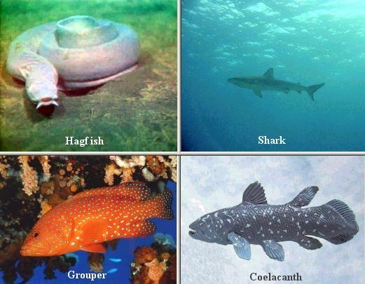 |
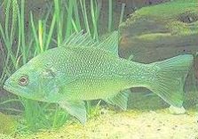
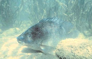
Figure 21, are a type of bony fish called ray-finned fishes. They have a swim bladder that aids them in changing their depth in the water. Species of fish that do not possess a swim bladder sink to the bottom if they stop swimming. "Ray-finned" refers to the fact that the fins are thin and are supported by bony rays. Another type of bony fish, called the lob-finned fishes, evolved into the amphibians. These fishes not only have fleshy appendages that could be adapted to land locomotion, they also have a lung that is used for respiration. The coelacanth (Figure 21), which exists today, is the only "living fossil" among the fishes. |
Figure 21 Fishes |
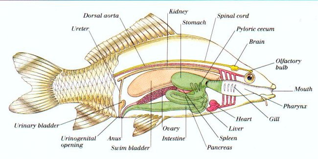
Figure 22 shows the internal anatomy of a common fish. |
 |
|
Figure 22 Fish Anatomy |
As the water passes over the gills, oxygen is absorbed by blood and carbon dioxide is given off. |
 |
|
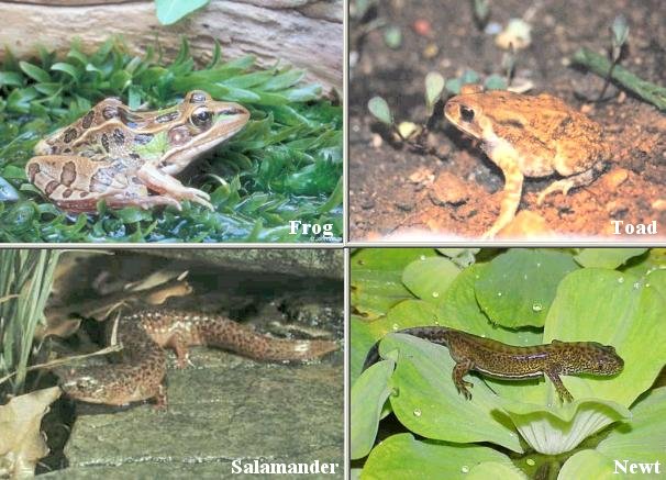
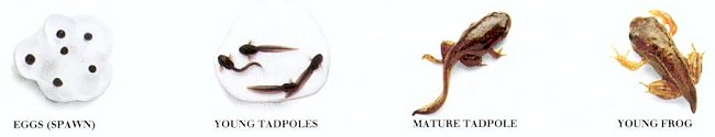
Figure 23 Amphibians |
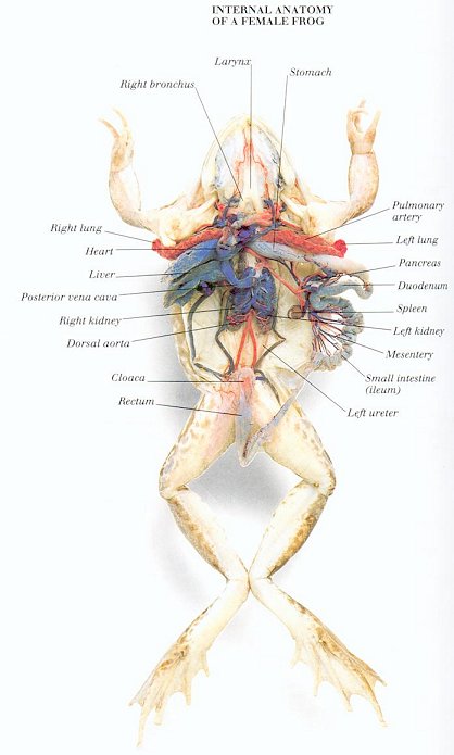 |
third eyelid, or nictitating membrane, may be drawn over the pulled-in eyeball. There is no external ear. Both eardrums, or tympanic membranes, are exposed. There is only one bone in the frog's middle ear. The human middle ear contains three bones (ossicles). As in man, semicircular canals help to maintain body balance.
|
Figure 24 Frog Anatomy |
 |
|
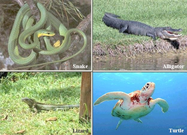
Figure 25 Reptiles |
by exposing themselves to the sun if they need warmth or by hiding in the shadows if they want to cool off. |
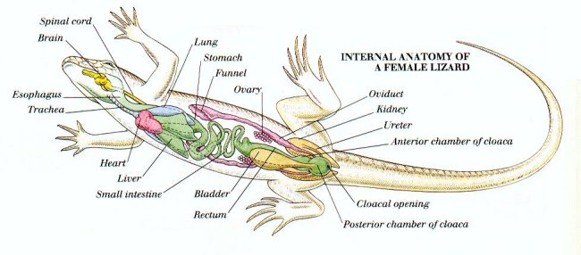 |
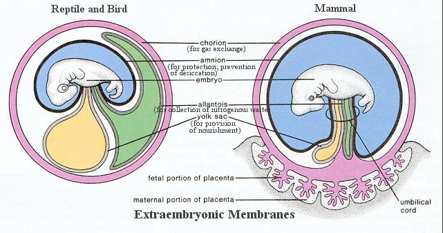
shelled egg made development on land possible and eliminated the need for a swimming-larva stage during development. It provides the developing embryo with oxygen, food, and water; it removes nitrogenous wastes; and it protects the embryo from drying out and from mechanical injury. This is accomplished by the presence of extraembryonic membranes. |
Figure 24 Lizard Anatomy |
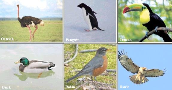 |
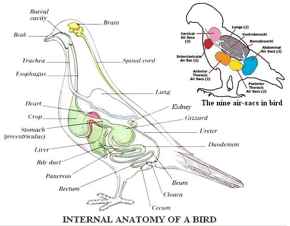
Birds are characterized by the presence of feathers, which are actually modified reptilian scales. There are many orders of birds, including birds that are flightless (ostrich), web footed (penguin), divers (loons), fish eaters (pelicans), waders (flamingos), broad billed (ducks), birds of prey (hawks), vegetarians (fowl), shorebirds (sandpipers), nocturnal (owl), small (hummingbirds), and songbirds, the most familiar of the birds. Some of them are showed in Figure 25. |
Figure 25 Birds |
Nearly every anatomical feature of a bird can be related to its ability to fly. Figure 26 shows the anatomy of a common bird. |
 |
|
Figure 26 Bird Anatomy |
sacs when the bird exhales - this means that the bird receives oxygen during inhalation and exhalation. Another benefit of air sacs is that the air-filled, hollow bones lighten the body and aid flying. |
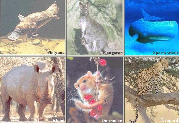 |
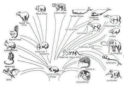
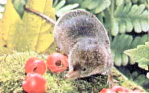
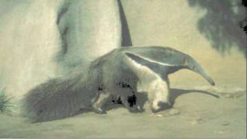
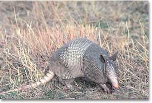
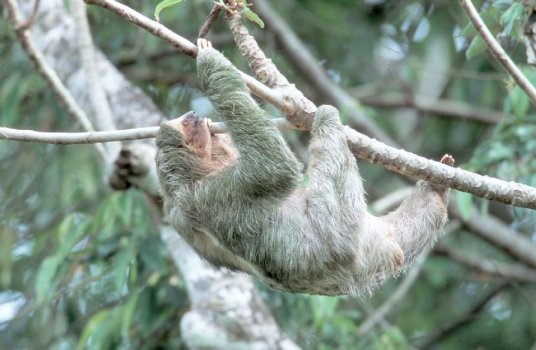
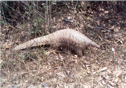
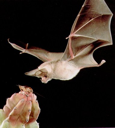
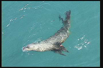
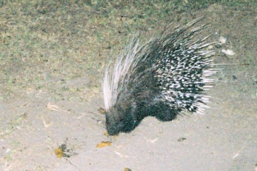
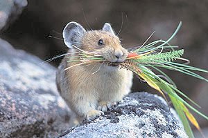
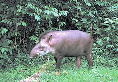
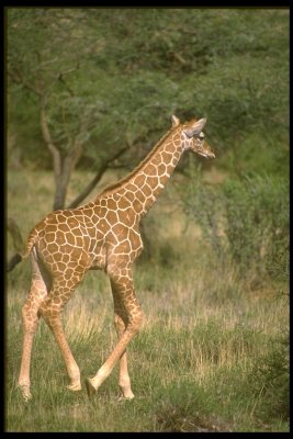
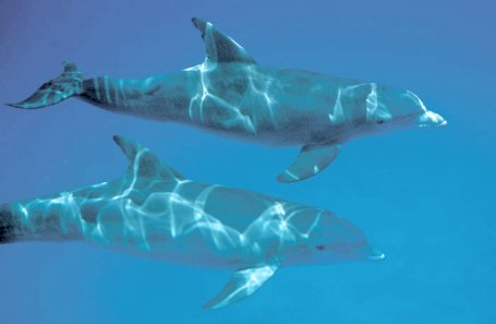
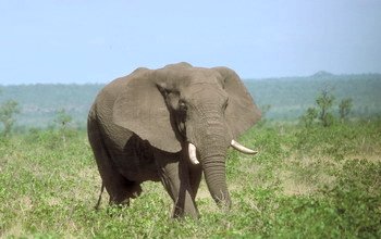
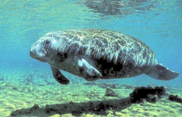
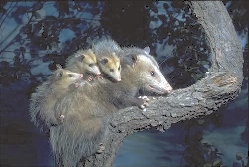
The chief characteristics of mammals are hair and mammary glands that produce milk to nourish the young. Human mammary glands are called breasts. Mammals are classified according to their means of reproduction: there are egg-laying mammals called monotremes such as the duck-billed platypus; mammals with pouches for immature embryos are the marsupials such as the kangaroos; while the placental mammals are the majority of living mammals. Figure 27 shows just a few of these animals. Table 02 lists the twelve orders of placental mammals. They are classified largely according to the mode of locomotion and how they get their food. Figure 28 illustrates a cat's anatomy, |
Figure 27 Mammals |
which is very similar to the human's. |
| Order | Examples | Characteristics |
|---|---|---|
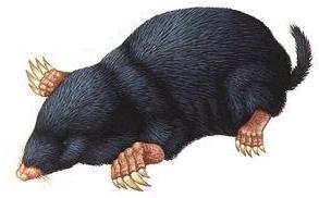
| Insectivora | Moles, shrews | Primitive; small, sharp-pointed teeth |
| Edentata | anteaters, armadillos, sloths | Primitive terrestrial mammal; few or no teeth; well developed claws |
| Pholidota | Pangolin | Medium size; large, plate-like scales; lack teeth, use powerful front claws and long tongues to reach ants or termite |
| Chiroptera | Bats | Digits support membranous wings |
| Carnivora | Dogs, bears, cats, sea lions | Long canine teeth; pointed teeth |
| Rodentia | Mice, rats, squirrels, beavers, porcupines | Incisor teeth grow continuously |
| Lagomorpha | Rabbits, hares, pikas | Chisel-like incisors; hind legs longer than front legs; herbivorous |
| Perissodactyla | Horses, zebras, tapirs, rhinoceroses | Large, long-legged, one or 3 toes, each with hoof; grinding teeth |
| Artiodactyla | Pigs, camels, buffalos, giraffes | Medium to large; 2/4 toes, each with hoof; many with antlers/horns |
| Cetacea | Whales, porpoises | Medium to very large; paddlelike forelimbs; hind limbs absent |
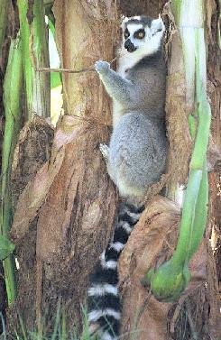
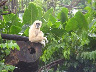
| Primates | Lemurs, monkeys, gibbons, chimpanzees, gorillas | Mostly tree dwelling; head freely movable on neck; 5 digits, usually with nails; thumbs and/or large toes usually opposable |
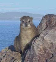
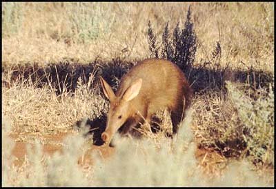
| Subungulate | Elephants, sirenians, hyraxes, aardvarks | Medium to very large; lack a clavicle, and have nails or hooves instead of claws. |
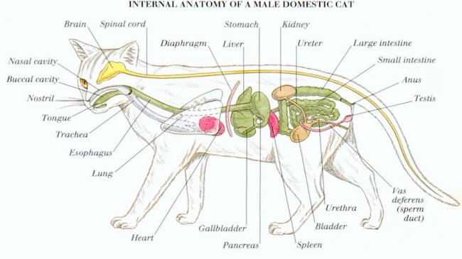 |
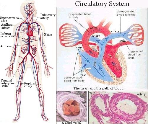
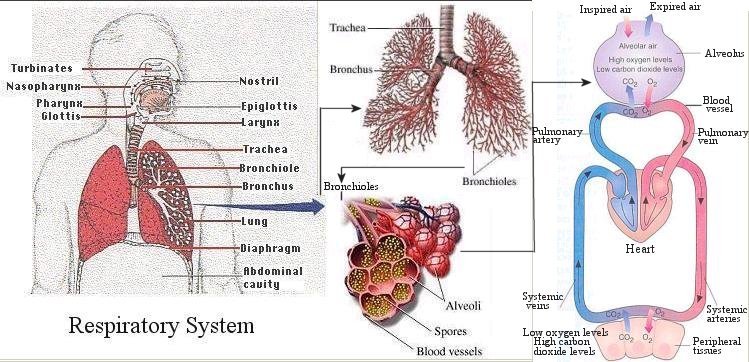
contract to some degree. As a result, air is sucked into and pushed out of the lungs through the trachea and the bronchial tubes or bronchi; these branch out and end in alveoli which are tiny sacs surrounded by capillaries filled with blood. Here oxygen from the air diffuses into the blood, where it is carried by hemoglobin. The deoxygenated blood from the heart reaches the lungs via the pulmonary artery and, after having been oxygenated, returns via the pulmonary veins. |
Figure 28 Cat Anatomy |
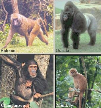 |
Humans are mammals in the order Primates. The first primates may have resembled today's tree shrews, rat-size animals with a snout, claws, and sharp front teeth. By 50 million years ago, however, primates had evolved characteristics suitable to move freely through the trees. The first primates were prosimians (meaning "premonkeys"). They are represented today by several types of animals, including the lemurs. Monkeys, along with apes and humans, are anthropoids. Monkeys evolved from the prosimians about 38 million years ago, when the weather was warm and vegetation was like that of a tropical rain forest. There are two types of monkeys: the New World monkeys such as the spider monkeys, which have long grasping tails and flat noses, and the Old World monkeys such as the baboons, which are now ground dwellers and lack such tails. Ape (gibbon, gorilla, and chimpanzee) evolved later. The human lineage split from that of the apes occurred about 5 - 10 million years ago in Africa. Figure 29 shows some of the primates. Figure 30 illustrates the chimpanzee anatomy, which is virtually identical to the human's. |
Figure 29 Primates |
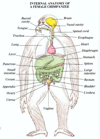 |
living. It is useful as an effective feeling and grasping mechanism to grab their insect prey, and to prevent them from falling and tumbling while moving through the trees. By far the most outstanding characteristic of primate evolution has been the enlargement of the brain among members of the order. Primate brains tend to be large, heavy in proportion to body weight, and very complex.
|
Figure 30 Chimp Anatomy |
![[view large image]](I10-82-Grasshopper.jpg)
![[view large image]](I10-82-Fishes.jpg)
![[view large image]](I10-82-Lizard.jpg)