 |
 |
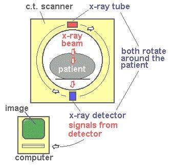
information than X rays used by themselves. CT scan also has the advantage of minimizing the amount of radiation exposure. Figure 1 is a schematic of the equipments with a rotating X rays source and detector. Before the scan is carried out, a contrast medium may be injected to make blood vessels, organs, or abnormalities show up more clearly; a drink of contrast medium may be given to highlight loops of intestine. |
Figure 1 CT Scan |
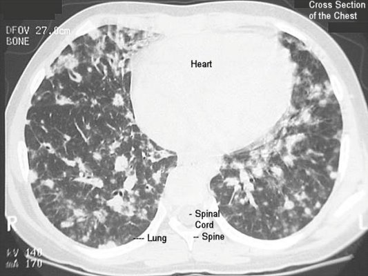
Figure 2 CT Image |
The amount of X rays absorbed by different tissues is recorded by the detector and transformed by a computer into an image. Figure 2 shows a CT cross-section of the chest. |
 |
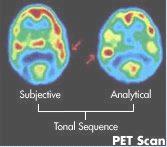 |
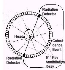
stream and are taken up in greater concentrations by areas of tissue that are more metabolically active. In the tissue, the substances emit positrons, which, in turn, release X-rays. It is the detection of these X-rays that actually forms the basis of PET scanning. By surrounding the patient with an array of detectors linked to a computer, the origin of the X-rays can be computed and a picture built of the distribution of the radio- active isotopes. Figure 4 is a sample of the PET brain scan. |
Figure 3 PET Scan |
Figure 4 PET Image |
 |
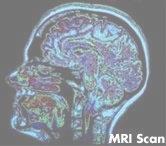 |
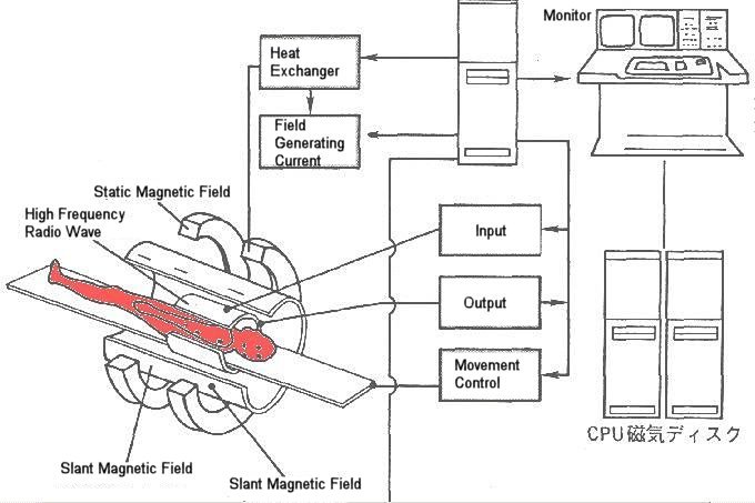
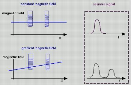
Magnetic field gradient is required to translates the signals into separate spatial locations. Magnetic coils in the machine detect these signals and a computer changes them into a cross-sectional or three dimensional image based on the strength of signal produced by different types of tissue. Tissues that contain a lot of hydrogen (such as fat) produce a bright image; those that contain little or no hydrogen (such as bone) appear dark. Figure 6 is a MRI image of the head. |
Figure 5 MRI Scan |
Figure 6 MRI Image |