|
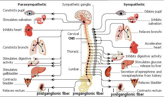
The human nervous system has three divisions: the central nervous system (CNS), the motor nervous system, and the sensory nervous system. The CNS consists of the brain and spinal cord. It acts as the central control region of the human nervous system, processing information and issuing commands. The autonomic (motor) nervous system is the command network the CNS uses to maintain the body's homeostasis. It automatically regulates heartbeat and controls muscle contractions in the walls of blood vessels, digestive, urinary, and reproductive tracts. It also carries messages that help stimulate glands to secrete tears, mucus, and digestive enzymes. |
[view large image] |
One division of the autonomic nervous system, called the sympathetic nervous system, dominates in times of stress. It controls the "fight or flight" reaction, increasing blood pressure, heart rate, breathing rate, and blood flow to the muscles. Another division, called the parasympathetic nervous system, has the opposite effect. It conserves energy by slowing the heartbeat and breathing rate, and by promoting digestion and elimination (of waste). Most glands, smooth muscles, and cardiac muscles constantly get inputs from both the sympathetic and parasympathetic systems. The CNS controls the activity by varying the ratio of the signals. Depending on which motor neurons are selected by the CNS, the net effect of the arriving signals will either stimulate or inhibit the organ. The above figure shows the various organs and actions, which are related to the two different divisions.
Motor fibers that govern involuntary responses, do not lead directly to the organs they innervate. Instead, they make their trips in two stages. The first set of fibers leads from the CNS to ganglia (which are collections of nerve cell bodies) that lie outside the CNS (the preganglionic fibers). At the ganglia the fibers form synaptic junctions with the dendrites of as many as twenty different cell bodies. The axons of these cell bodies form a second set of fibers, the postganglionic fibers. It is these postganglionic fibers that lead to the organs.
The chief ganglia involved in the autonomic nervous system form two lines running down either side of the spinal column. They are outside the bony vertebrae. These two lines of ganglia outside the column resemble a pair of long beaded cords. At the lower end, the two cords join and finish in a single central stretch. These lines of ganglia are sometimes called the sympathetic trunks (used by the sympathetic nervous system). Not all ganglia are located in the sympathetic trunks. Some are not; and it is possible for a preganglionic fiber to go right through, making no synaptic junction there at all, joining instead with ganglia located in front of the vertebrae. For the parasympathetic nervous system, some of the ganglia separating the preganglionic fibers from the postganglionic fibers are actually located within the organ the nerve is servicing. In that case, the preganglionic fiber runs almost the full length of the total track, whereas the postganglionic fiber is at most just a few millimeters long.
The splanchnic nerves, which originate from some of the thoracic nerves, have their preganglionic fibers ending in a mass of ganglia lying just behind the stomach. It represents the largest mass of nerve cells that is not within the CNS and is sometimes called the "abdominal brain". It is a vital place to be protected during boxing.